Hemodynamic Monitoring
Hemodynamic Monitoring. Charles E. Smith, MD Professor of Anesthesia Director, Cardiothoracic Anesthesia MetroHealth Medical Center Case Western Reserve University Cleveland, Ohio Email: csmith@metrohealth.org. Definition of Monitoring.
Share Presentation
Embed Code
Link
Download Presentation

staciae + Follow
Download Presentation
Hemodynamic Monitoring
An Image/Link below is provided (as is) to download presentation Download Policy: Content on the Website is provided to you AS IS for your information and personal use and may not be sold / licensed / shared on other websites without getting consent from its author. Content is provided to you AS IS for your information and personal use only. Download presentation by click this link. While downloading, if for some reason you are not able to download a presentation, the publisher may have deleted the file from their server. During download, if you can't get a presentation, the file might be deleted by the publisher.
Presentation Transcript
- Hemodynamic Monitoring Charles E. Smith, MD Professor of Anesthesia Director, Cardiothoracic Anesthesia MetroHealth Medical Center Case Western Reserve University Cleveland, Ohio Email: csmith@metrohealth.org
- Siegel JH et al: Trauma: Emergency Surgery + Critical Care, 1987:201-284
- Definition of Monitoring • Continuous or repeated observation + vigilance in order to maintain homeostasis • ASA Standards: I. Qualified personnel II. Oxygenation: SaO2, FiO2 III. Ventilation: ETCO2, stethoscope, disconnect alarm IV. Circulation: BP, pulse, ECG Other monitors: T, Paw, Vt, ABG
- Objectives • Arterial line • Systolic pressure variation • Central venous pressure • Pulmonary artery catheterization • Cardiac output • Mixed venous oxygen
- Basic Concepts • BP = CO x SVR • CO = SV x HR • DO2= (CO x CaO2x 10) + (PaO2x 0.003) • CaO2 = Hg x 1.39 x O2 sat; or CaO2= Hct/2 Assume CO 5 L/min, 100% sat • Hct 40 CaO2 20 CO 5 DO2 1000 • Hct 30 CaO2 15 CO 5 DO2 750 • Hct 20 CaO2 10 CO 5 DO2 500
- Arterial Line • Indications: • Rapid moment to moment BP changes • Frequent blood sampling • Circulatory therapies: bypass, IABP, vasoactive drugs, deliberate hypotension • Failure of indirect BP: burns, morbid obesity • Pulse contour analysis: SPV, SV
- Radial Artery Cannulation • Technically easy • Good collateral circulation of hand • Complications uncommon except: • vasospastic disease • prolonged shock • high-dose vasopressors • prolonged cannulation
- Alternative Sites • Brachial: • Use longer catheter to traverse elbow joint • Postop keep arm extended • Collateral circulation not as good as hand • Femoral: • Use guide-wire technique • Puncture femoral artery below inguinal ligament (easier to compress, if required)
- Pulsus Paradoxus • Exaggerated inspiratory fall in systolic BP during spontaneous ventilation, > 10-12 mmHg • Cardiac tamponade, severe asthma
- Systolic Pressure Variation • Difference between maximal + minimal values of systolic BP during PPV • down: ~ 5 mm Hg due to venous return • SPV > 15 mm Hg, or down > 15 mm Hg: • highly predictive of hypovolemia Marik: Anaesth Intensive Care 1993;21:405. Coriat: Anesth Analg 1994;78:46
- Gardner, in Critical Care, 3rd ed. Civetta. 1997, p 851
- Pulse Contour Analysis • 1. Transform BP waveform into volume – time waveform • 2. Derive uncalibrated SV • SV x HR = CO • 3. May calibrate using Li indicator [LidCO] or assume initial SV based on known EF from echo • Assumptions: • PPV induces cyclical changes in SV • Changes in SV results in cyclical fluctuation of BP or SPV Linton R: 1997, 1998, 2000
- PulseCO SPV + SV • Predicts SV in response to volume after cardiac surgery + in ICU[Reuter: BJA 2002; 88:124; Michard: Chest 2002; 121:2000] • Similar estimates of preload v. echo during hemorrhage[Preisman: BJA 2002; 88: 716] • Helpful in dx of hypovolemia after blast injury [Weiss: J Clin Anesth 1999; 11:132]
- Pitfalls with SPV + SV • Inaccurate if • AI • IABP • Problems if • pronounced peripheral arterial vasoconstriction • damped art line • arrhythmias
- Central Venous Line • Indications: • CVP monitoring • Advanced CV disease + major operation • Secure vascular access for drugs: TLC • Secure access for fluids: introducer sheath • Aspiration of entrained air: sitting craniotomies • Inadequate peripheral IV access • Pacer, Swan Ganz
- Central Venous Line: RIJ • IJ vein lies in groove between sternal + clavicular heads of sternocleidomastoid muscle • IJ vein is lateral + slightly anterior to carotid • Aseptic technique, head down • Insert needle towards ipsilateral nipple • Seldinger method: 22 G finder; 18 G needle, guidewire, scalpel blade, dilator + catheter • Observe ECG + maintain control of guide-wire • Ultrasound guidance; CXR post insertion
- Advantages of RIJ • Consistent, predictable anatomic location • Readily identifiable landmarks • Short straight course to SVC • Easy intraop access for anesthesiologist at patient’s head • High success rate, 90-99%
- Types of Central Catheters • Variety of lengths, gauges, composition + lumens depending on purpose • Introducer sheath (8-8.5 Fr): • Permits rapid fluid/blood infusion or Swan • Trauma triple-lumen (12 Fr): • Rapid infusion via 12 g x 2; 16 g for CVP monitoring • MAC 2: (9 Fr): • Rapid infusion via distal port; 12 g for CVP • Also allows for Swan insertion • More septations + stiffer plastic
- Alternative Sites • Subclavian: • Easier to insert v. IJ if c-spine precautions • Better patient comfort v. IJ • Risk of pneumo- 2% • External jugular: • Easy to cannulate if visible, no risk of pneumo • 20%: cannot access central circulation • Double cannulation of same vein (RIJ) • Serious complications: vein avulsion, catheter entanglement, catheter fracture
- CVP Monitoring • Reflects pressure at junction of vena cava + RA • CVP is driving force for filling RA + RV • CVP provides estimate of: • Intravascular blood volume • RV preload • Trends in CVP are very useful • Measure at end-expiration • Zero at mid-axillary line
- Zero @ Mid-Axillary Line
- CVP Waveform Components Mark JB, CV Monitoring, in Miller 5th Edition, 2000, pg 1153
- Pulmonary Artery Catheter • Introduced by Swan + Ganz in 1970 • Allows accurate bedside measurement of important clinical variables: CO, PAP, PCWP, CVP to estimate LV filling volume, + guide fluid / vasoactive drug therapy • Discloses pertinent CV data that cannot be accurately predicted from standard signs + symptoms
- PAC Waveforms
- Indications: ASA Task Force • Original practice guidelines for PAC- 1993; updated 2003 [Anesthesiology 2003;99:988] • High risk patient with severe cardiopulmonary disease • Intended surgery places patient at risk because of magnitude or extent of operation • Practice setting suitable for PAC monitoring: MD familiarity, ICU, nursing • PAC Education Project: www.pacep.org • web based resource for learning how to use PAC Roizen et al: Anesthesiology 1993;78:380. ASA Newsletter, Aug 2002;66(8):7
- PAC and Outcome • Early use of PAC to optimize volume status + tissue perfusion may be beneficial • PAC is only a monitor. It cannot improve outcome if disease has progressed too far, or if intervention based on PAC is unsuccessful or detrimental • Many confounding factors: learning bias, skill, knowledge, usage patterns, medical v. surgical illness Connors: JAMA 1996;276:916. Mark JB: in Anesthesia 5th Ed. Miller. 2000: pp 1178-80
- PAC: Complications • Minor in 50%, e.g., arrhythmias • Transient RBBB- 0.9-5% • External pacer if pre-existing LBBB • Misinformation • Serious: 0.1-0.5%: knotting, pulmonary infarction, PA rupture (e.g., overwedge), endocarditis, structural heart damage • Death: 0.016% Mark JB, in Anesthesia 5th Edition. Miller 2000, pg 1117-1206
- Problems Estimating LV Preload
- Cardiac Output • Important feature of PAC • Allows calculation of DO2 • Thermodilution: inject fixed volume, 10 ml, (of room temp or iced D5W) into CVP port at end-expiration + measure resulting change in blood temp at distal thermistor • CO inversely proportional to area under curve
- Cardiac Output: Technical Problems • Variations in respiration: • Use average of 3 measures • Blood clot over thermistor tip: inaccurate temp • Shunts: LV + RV outputs unequal, CO invalid • TR: recirculation of thermal signal, CO invalid • Computation constants: • Varies for each PAC, check package insert + manually enter
- Continuous Mixed Venous Oximetry • Fick Equation • VO2 = CO [CaO2 - CvO2] • CvO2 ~ SvO2 b/c most O2 in blood bound to Hg • If O2 sat, VO2 + Hg remain constant, SvO2 is indirect indicator of CO • Can be measured using oximetric Swan or CVP, or send blood gas from PA / CVP • Normal SvO2 ~ 65% [60-75]
- Mixed Venous Oximetry • ↑ SvO2 [> 75%] • Wedged PAC: reflects LAP saturation • Low VO2: hypothermia, general anesthesia, NMB • Unable to extract O2 : cyanide, Carbon monoxide • High CO: sepsis, burns, L→ R shunt AV fistulas
- Mixed Venous Oximetry • ↓ SvO2 [ < 60%] • ↓ Hg- bleeding, shock • ↑ VO2: fever, agitation, thyrotoxic, shivering • ↓ SaO2 : hypoxia, resp distress • ↓ CO: MI, CHF, hypovolemia
- Summary • Invasive monitoring routinely performed • Permits improved understanding of BP, blood flow, + CV function • Allows timely detection of hemodynamic events + initiation of treatment • Requires correct technique + interpretation • Complications occur from variety of reasons • Risk: benefit ratio usually favorable in critically ill patients

hemodynamic monitoring
OBJECTIVE . 10/1/2011. 2. 1. Describe the three attributes of circulating blood and their relationships.2. Identify types of clients in which hemodynamic monitoring would be indicated.3. Describe the types of catheters used for hemodynamic monitoring.4. Discuss the normal and abnormal values obtained through hemodynamic monitoring as they relate to specific client situations.5. List the potential complications in use of hemodynamic monitoring devices. 6. Explain nurs9444
1.5k views • 26 slides

Hemodynamic monitoring
. . . Indications for pulmonary artery catheterization in the ICU:Establish diagnosis of shock and/or respiratory failureGuide therapy of shock and/or respiratory failureBy improving oxygen delivery. . Oxygen delivery = CaO2 x COCardiac output = HR x SVSV is determined by:Preload (end-diastolic volume)Cardiac contractilityAfterload .
1.48k views • 69 slides

Hemodynamic Monitoring
Hemodynamic Monitoring. By Nancy Jenkins RN,MSN. What is Hemodynamic Monitoring and why do it?. It is measuring the pressures in the heart It allows us to see inside the heart and adjust volume as needed. Comparing Hemodynamics to IV pump. Fluid =preload
4.88k views • 62 slides
MODULE F – HEMODYNAMIC MONITORING
MODULE F – HEMODYNAMIC MONITORING. Topics to be Covered. Cardiac Output Determinants of Stroke Volume Hemodynamic Measurements Pulmonary Artery Catheterization Control of Blood Pressure Heart Failure. Cardiac Measurements.
1.62k views • 41 slides
Advance in Hemodynamic Monitoring
Advance in Hemodynamic Monitoring. By Dr H P Shum. Outline. Introductions What we have previously – A line / CVC/ PAC Advance techniques for haemodynamic monitoring. Introductions.
2.8k views • 72 slides

Hemodynamic Monitoring
Hemodynamic Monitoring. Charles E. Smith, MD Professor of Anesthesia Director, Cardiothoracic Anesthesia MetroHealth Medical Center Case Western Reserve University Cleveland, Ohio Email: csmith@metrohealth.org. Definition of Monitoring.
2.29k views • 44 slides

HEMODYNAMIC MONITORING
mlr/2007. OBJECTIVES. The student will reviewcardiac and pulmonary considerations for invasive monitoringProcedural considerations for invasive monitoringWaveform identification related to invasive monitors. . mlr/2007. EVALUATING THE PATIENT A REVIEW. PULMONARYBreath soundsLevel of mentatio
1.56k views • 89 slides
Hemodynamic Monitoring and Transthoracic Lines
Hemodynamic Monitoring and Transthoracic Lines. Deb Updegraff RN, CNS Lucille Packard Children’s Hospital Pat Hock RN, Nurse Educator. Winnie Yung , CNS. Infants and children undergoing open heart surgery may require intracardiac monitoring.
1.14k views • 21 slides
Hemodynamic Monitoring in the CCU
Hemodynamic Monitoring in the CCU. Edward G. Hamaty Jr., D.O. FACCP, FACOI. Waveform Review. Left Ventricular Pressure. Normal left ventricular pressures are: Systolic 100 to 140 mm of mercury End-diastolic 3 to 12 mm of mercury
2.11k views • 170 slides

HEMODYNAMIC MONITORING
HEMODYNAMIC MONITORING. Martha Richter, MSN, CRNA. OBJECTIVES. The student will review cardiac and pulmonary considerations for invasive monitoring Procedural considerations for invasive monitoring Waveform identification related to invasive monitors. EVALUATING THE PATIENT – A REVIEW.
1.67k views • 89 slides
Hemodynamic monitoring
Hemodynamic monitoring. Magdy M Khalil, MD, EDIC. Tissue perfusion. Oxygen delivery = CO x arterial oxygen content CO = (SV x HR) x <(Hb x 1.39 x SaO 2 ) + (0.003 x PaO 2 )>Arterial pressure (AP) = CO x SVR. Diagnosis of tissue malperfusion. Clinical assessment Basic monitoring
970 views • 18 slides
Hemodynamic Monitoring for the Respiratory Therapist
Hemodynamic Monitoring for the Respiratory Therapist. Jane Reynolds, MS, RN, RRT. Definition of terms. Preload – amount of blood in the ventricle before contraction – E nd d iastolic v olume EDV determines the amount of ‘stretch’ that is placed on the myocardial muscle
729 views • 53 slides

Hemodynamic Monitoring
Hemodynamic Monitoring. By Nancy Jenkins RN,MSN. What is Hemodynamic Monitoring?. It is measuring the pressures in the heart. Hemodynamic Monitoring. Baseline data obtained (low cardiac output) General appearance Level of consciousness Skin color/temperature Vital signs
979 views • 37 slides

Hemodynamic Monitoring
Hemodynamic Monitoring. Hemodynamics?. Hemo : blood Dynamics: movement Hemodynamics: movement of blood The measurement of the force (pressure) exerted by the blood as it moves through blood vessels or heart chambers during systole and diastole
2.59k views • 103 slides

Hemodynamic Monitoring in Sepsis
Hemodynamic Monitoring in Sepsis. Shao-Hsuan Hsia, MD Pediatric Critical Care Medicine Chang Gung Children’s Hospital. Pathophysiology of septic shock Cardiac output monitoring Tissue perfusion monitoring Demand-supply balance. Definition of sepsis. Infection Bacteremia
1.18k views • 55 slides
Hemodynamic Monitoring
Hemodynamic Monitoring. John Nation RN, MSN Thanks to Nancy Jenkins. What is Hemodynamic Monitoring?. It is measuring the pressures in the heart. Hemodynamic Monitoring. Baseline data obtained General appearance Level of consciousness Skin color/temperature Vital signs
2.29k views • 32 slides

INVASIVE HEMODYNAMIC MONITORING
INVASIVE HEMODYNAMIC MONITORING. Presentation by Donna Cohen, BSN, RN Heart and Vascular Center Medical University of South Carolina February 2006. INVASIVE HEMODYNAMIC MONITORING. Objectives.
2.26k views • 39 slides
Effect of pulmonary hypertension on hemodynamic monitoring
Effect of pulmonary hypertension on hemodynamic monitoring. Richard Siebert Physician / critical care fellow Dept. Critical Care : Steve Biko Academic Hospital/lcm hospital. Why hemodynamic monitoring in ICU?. We are interested in maintaining and optimising organ perfusion.
505 views • 17 slides
Hemodynamic Monitoring Part I (ABP, CVP, Ao)
Hemodynamic Monitoring Part I (ABP, CVP, Ao). MICU Competencies 2006-2007. What is Hemodynamic Monitoring?. Non-invasive = clinical assessment & NBP Direct measurement of arterial pressure Invasive hemodynamic monitoring. Noninvasive BP Heart Rate, pulses Mental Status Mottling (absent).
851 views • 35 slides
Hemodynamic Monitoring
Hemodynamic Monitoring. Khaled O. Hadeli 12/7/99. DO2 = CO x 13.4 x Hb x SaO2 DO2 = (SV x HR) 13.4 x Hb x SaO2. MR. RVF. Hypovolemic shock. Acute bronchospasm. Busy Tracing. Cardiac performance CO/CI CVP/RAP/RVP/PAP/ Pcwp RVEF SVR/PVR. O2 transport parameters
807 views • 45 slides
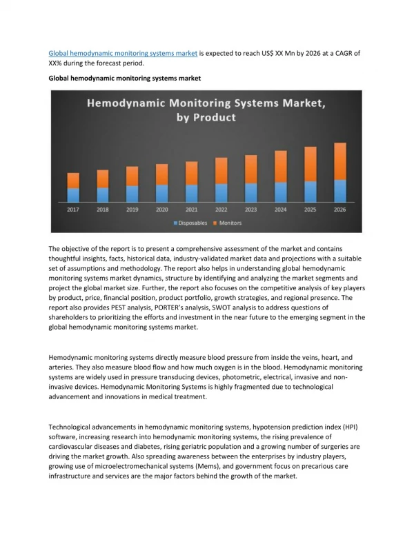
Global Hemodynamic Monitoring Systems Market
Global hemodynamic monitoring systems market is expected to reach US$ XX Mn by 2026 at a CAGR of XX% during the forecast period.
Chapter 9 Hemodynamic Monitoring
Chapter 9 Hemodynamic Monitoring. Indications for Hemodynamic Monitoring. Assesses cardiac function and evaluates effectiveness of therapy Cardiogenic shock Severe heart failure Sepsis or septic shock Multiple organ system dysfunction (MODS) Acute respiratory distress syndrome (ARDS)
552 views • 43 slides
